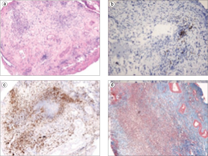
Figure 2.
Brain biopsy findings suggesting a diagnosis of hypertrophic pachymeningitis. (a) Hematoxylin and eosin stain showing edematous cortex with attached chronically inflamed fibrous dura mater. (b) CD20 stain demonstrating scattered B lymphocytes. (c) CD68 stain demonstrating an abundance of macrophages. (d) Trichrome stain highlighting blue-staining dense fibrosis. Stains for organisms were negative, and IgG4 highlighted rare cells.

