Figure 7.
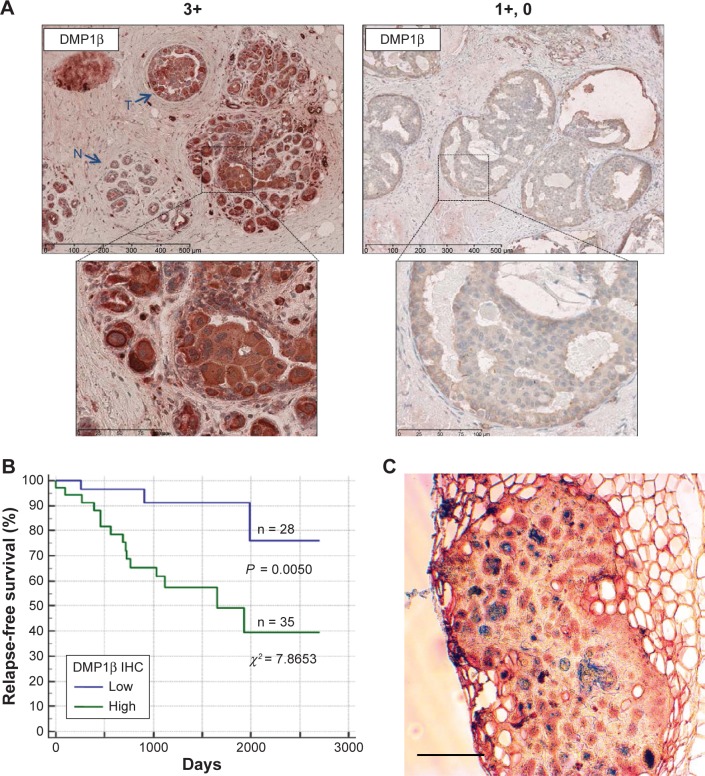
DMP1β IHC in human BC. (A) Representative images of DMP1β IHC staining from two BC patients (Patient #1: high DMP1β expression; Patient #2: low DMP1β expression).59 A total of 63 human breast tumors were stained with DMP1β-specific antibody, RAB. DMP1β staining was significantly higher in the tumor tissues in Patient #1 compared to surrounding normal tissues. The scale bar indicates 100 µm. (B) A Kaplan–Meier RFS curve was graphed based on high versus low DMP1β protein intensity.59 Patients with significantly higher DMP1β (high) staining in tumors compared to the surrounding normal tissue had significantly shorter relapse than the those with tumors that show undetectable DMP1β (low) in their tumor tissue (P = 0.0050; c2 = 7.8653). (C) Mammary tumor tissue from an MMTV-DMP1βV5His female mouse doubly stained for DMP1β (peroxidase; red) and cytokeratin 14 (alkaline phosphatase; blue). The majority of tumor cells were positive for both proteins, suggesting transdifferentiation of mammary tumor cells to adenosquamous carcinoma. The scale bar indicates 100 µm.

