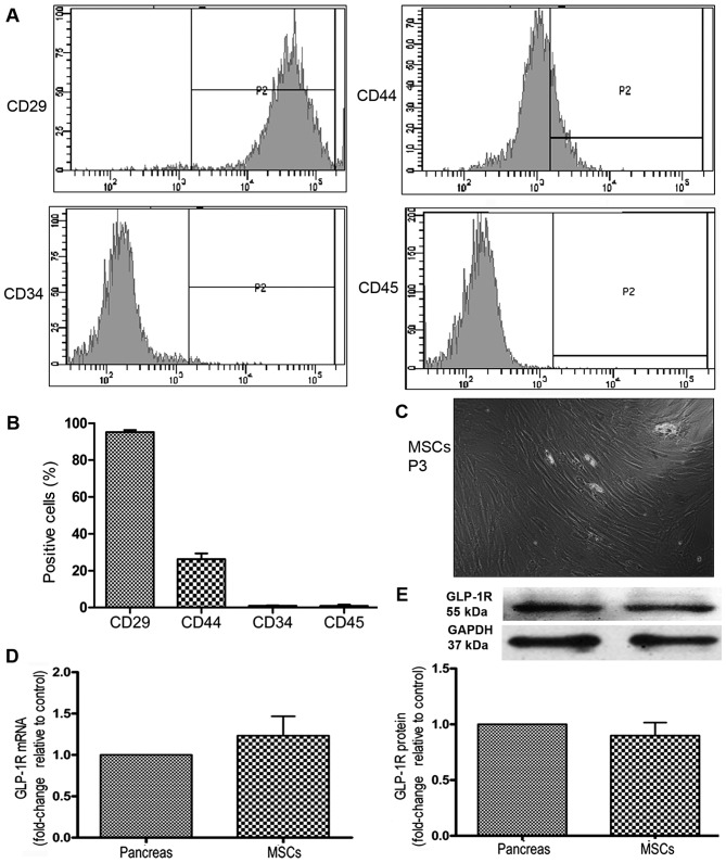
Figure 1.
Characterization of mesenchymal stem cells (MSCs). MSC cell surface markers were detected by FACS. (A) Representative histograms of immuophenotypic assay for CD29, CD44, CD34 and CD45. (B) Quantitative analysis of cell surface marker expression. (C) Image of MSCs at passage 3 (P3) captured by microscopy at a magnification of x100. (D) The mRNA expression of glucagon-like peptide-1 receptor (GLP-1R) on MSCs was detected by RT-qPCR. Its expression on pancreas served as a positive control. (E) The protein expression of GLP-1R on MSCs was detected by western blot analysis. Its expression on pancreas was served as a positive control.

