Figure 2.
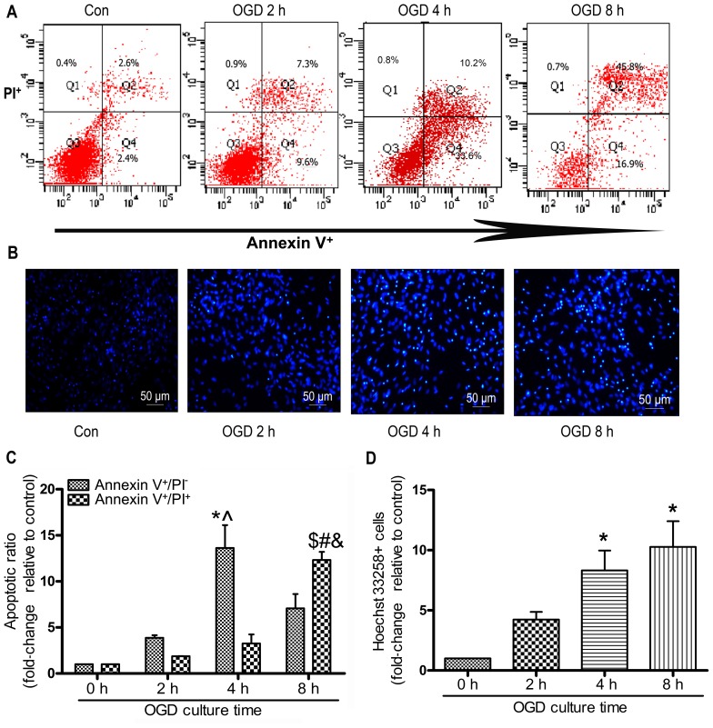
Oxygen and glucose deprivation (OGD) induces the apoptosis of mesenchymal stem cells (MSCs). MSCs were subjected to OGD conditions for the indicated periods of times, and the apoptotic ratio was detected. Apoptosis was assessed by Annexin V/propidium iodide (PI) double staining and FACS. (A) Representative FACS plots. (B) The nuclei of the MSCs were dyed with Hoechst 33258 and representative images of Hoechst 33258-stained cells were obtained under a fluorescence microscope; each experiment was performed 3 times, (C) Quantitative analysis of percentage of early-stage apoptotic cells (Annexin V+/PI−) and late-stage apoptotic (Annexin V+/PI+) cells, each column represents the means ± SD of 3 independent experiments. Annexin V+/PI− cells: *P<0.05 vs. 0 h group; ^P<0.05 vs. 8 h group; Annexin V+/PI+ cells: $P<0.05 vs. 0 h group; #P<0.05 vs. 2 h group; &P<0.05 vs. 4 h group. (D) Quantitative analysis of Hoechst 33258 stained-cells, the cells were shown at magnification of x100, each column represents mean ± SD of 3 independent experiments. *P<0.05 vs. 0 h group.

