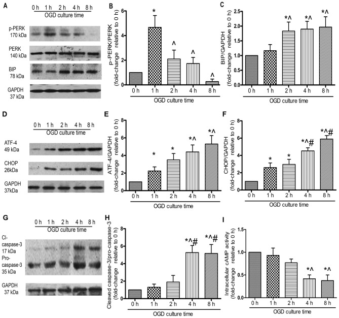
Figure 3.
Endoplasmic reticulum (ER) stress is involved in the oxygen and glucose deprivation (OGD)-mediated apoptosis of mesenchymal stem cells (MSCs). MSCs were incubated under OGD conditions for the indicated periods of time, and the protein was extracted and quantified by western blot (WB) analysis. All experiments were performed in triplicate. (A) Representative western blots of phosphorylated (p-)PERK, PERK, BIP and the loading control, GAPDH. Relative quantity of (B) p-PERK was estimated as fold-changes relative to PERK, (C) BIP was estimated as fold-changes relative to GAPDH. (D) Representative western blots of ATF-4 and CHOP. Quantitative analysis of (E) ATF-4 and (F) CHOP was estimated as fold-changes relative to GAPDH. (G) Representative western blots of cleaved caspase-3 and pro-caspase-3. (H) The expression of cleaved caspase-3 was estimated as fold-changes relative to pro-caspase-3. (I) MSCs were subjected to OGD conditions for the indicated periods of time and intracellular cAMP activity was assessed by ELISA, each column represents the means ± SD of 3 independent experiments. *P<0.05 vs. 0 h; ^P<0.05 vs. 1 h; #P<0.05 vs. 2 h.

