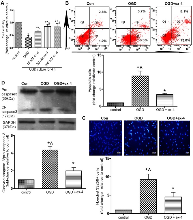
Figure 4.
Exendin-4 (ex-4) protects mesenchymal stem cells (MSCs) from oxygen and glucose deprivation (OGD)-induced apoptosis. MSCs were pre-treated with ex-4 for 12 h and subsequently incubated under OGD conditions for 4 h. All experiments were conducted in triplicate. (A) Cell viability was detected by an MTT assay. *P<0.05 vs. control group; ^P<0.05 vs. OGD group; #P<0.05 vs. 10 nM ex-4 group. Apoptosis was detected by Annexin V/propidium iodide (PI) staining and FACS. (B) Representative FACS plots and quantitative analysis of the apoptotic ratio. The apoptotic ratio was the percentage summation of early-stage apoptotic cells (Q4) and late-stage apoptotic (Q2) cells. (C) Representative images of Hoechst 33258-stained cells and quantitative analysis of the ratio of Hoechst 33258-positive cells. The cells were shown at magnification of x100. (D) The expression of cleaved caspase-3 and pro-caspase-3 detected by western blot analysis. For (B–D) *P<0.05 vs. control group; ^P<0.05 vs. OGD + ex-4 group.

