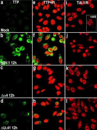
FIG. 4.
Localization of TTP and TIA-1 in HEp-2 cells infected with HSV-1. HEp-2 cells were mock infected (a, e, and i) or exposed to 10 PFU of HSV-1(F) (b, f, and j) or the Δα4 (c, g, and k) or ΔUL41 (d, h, and l) mutant virus per cell and fixed by paraformaldehyde and then methanol 12 h after infection. The cells were labeled with an anti-TTP antibody, detected with a FITC-labeled secondary antibody (green), and counterstained with propidium iodide (PI; red), or they were labeled with anti-TIA-1/TIAR antibodies and detected with an Alexa fluor 594-labeled secondary antibody (red). Immunostaining of the monolayers was evaluated by confocal microscopy. Cells were exposed to 0.5 mM arsenite for 30 min.

