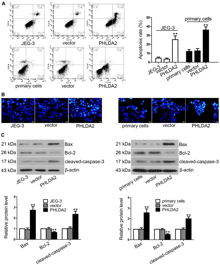
Figure 3.
Pleckstrin homology-like domain, family A, member 2 (PHLDA2) overexpression promotes cell apoptosis. (A) After infection with lentiviruses for 24 h, the apoptosis rate of JEG-3 cells and primary trophoblasts induced by PHLDA2 was measured using flow cytometry. (B) Apoptotic morphological changes in the JEG-3 and primary cells were detected by Hoechst staining and measured by fluorescence microscopy. Scale bar, 20 µm. (C) Proteins were extracted from the cells in each group and the concentrations were determined by BCA assay. Cleaved-caspase-3, Bax and Bcl-2 expression were analyzed by western blot analysis and normalized to β-actin expression. **P<0.01, compared with the control vector groups.

