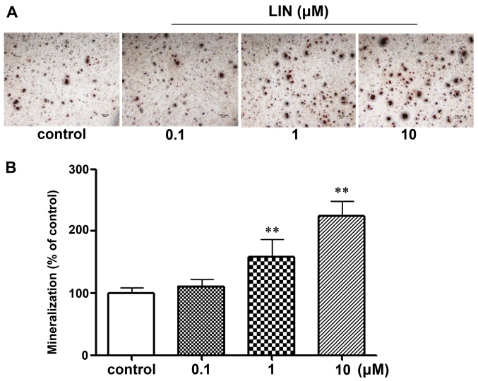
Figure 4.
Effect of linarin (LIN) on mineralization of MC3T3-E1 cells. (A) After treatment with various concentrations of LIN for 21 days, the mineral nodule formation was assessed by Alizarin red S dye (×4). Scale bar, 100 µm. (B) The bound, stained cells were washed with cetylpyridinium chloride and quantified using a Bio-Rad ELISA reader. LIN increased the mineralization of MC3T3-E1 cells. Data are represented as the means ± SD of three independent experiments. **P<0.01 as compared with control.

