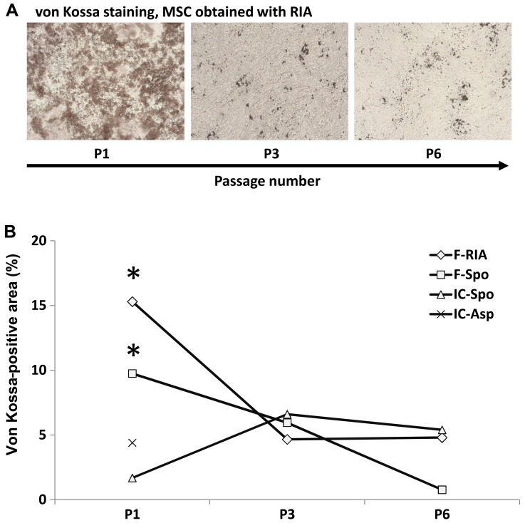
Figure 5.
Decline in calcium deposition with the increasing passage number of mesenchymal stem cells (MSCs). (A) Representative images of von Kossa staining of osteogenically differentiated MSCs of passage 1, 3 and 6 are shown (original magnification ×50). (B) Evaluation of the von Kossa staining. A significant decline in the von Kossa-positive areas was noted between MSCs of the 1st and MSCs of the 6th culture passages. MSCs were obtained from the femur using the Reamer/Irrigator/Aspirator (RIA) technique (diamond), from the femur using a spoon (square), from the iliac crest using a spoon (triangle) or from the iliac crest by fine-needle aspiratoin (x). Only data of the 1st culture passage were available for the MSCs obtained from the iliac crest. *p<0.05 culture passage 1 vs. culture passage 6.

