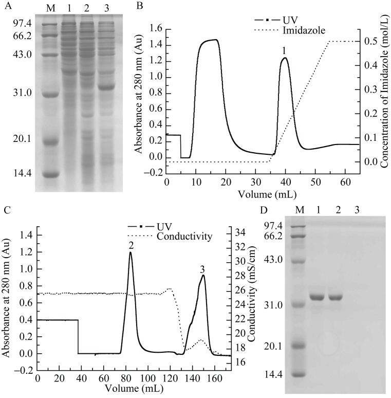
Figure 4.
Prokaryotic expression, purification and refolding of rLcIL-1β A: Bacterial lysates were electrophoresed on 12% SDS-PAGE gels. Lane M: protein marker; 1: E. coli BL21 (DE3) transformed with pET28a after IPTG induction; 2: E. coli BL21 (DE3) transformed with pET28a-LcIL-1β before IPTG induction; 3: E. coli BL21 (DE3) transformed with pET28a-LcIL-1β after IPTG induction. B: His affinity chromatography purification of rLcIL-1β using His TrapTM FF Crude column. C: Refolding of rLcIL-1β using urea gradient gel filtration on a Superdex 75 column. D: SDS-PAGE analysis of peaks in B and C. Lane M: protein marker; Lane 1: peak 1; 2: peak 2; 3: peak 3.

