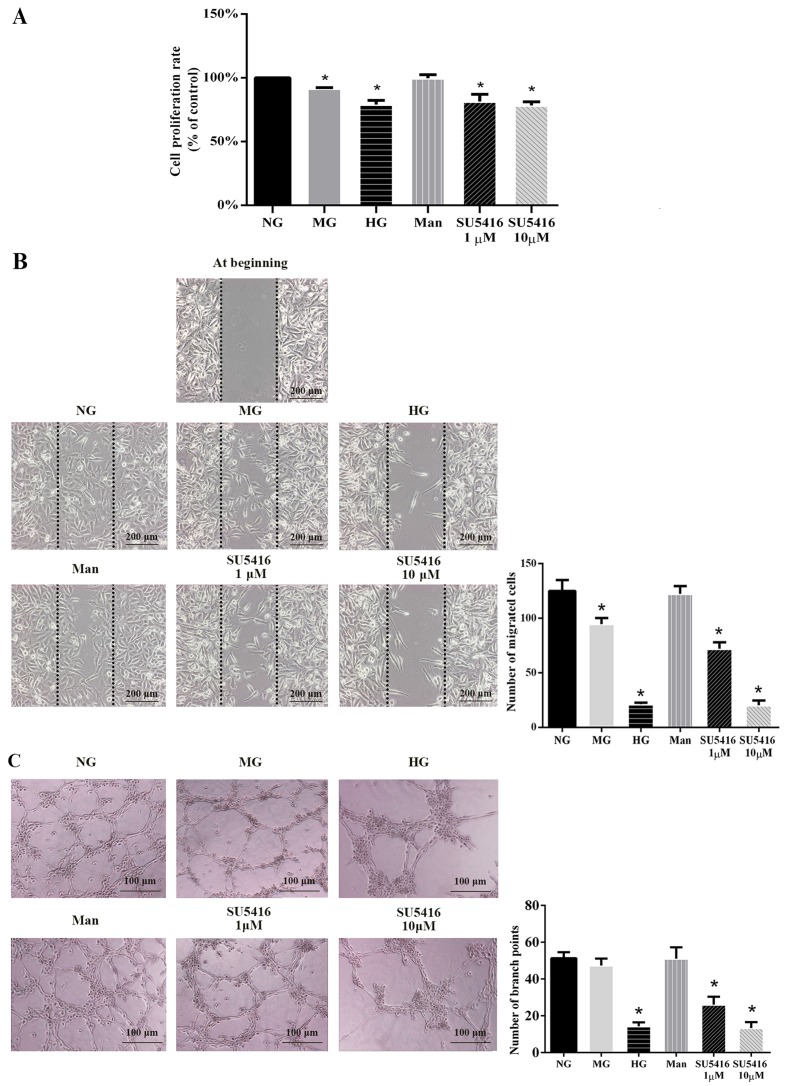
Figure 1.
Inhibition of cell proliferation, migration and tube formation in human umbilical vein endothelial cells (HUVECs) exposed to different concentrations of glucose [NG (normal glucose), MG (medium glucose) and HG (high glucose)], mannitol (Man, for osmotic control) and SU5416 (1 and 10 µM). (A) Cells of the six groups were cultured as indicated. Cell viability was determined by MTT assay. Results were presented as the cell proliferation rate described in Materials and methods. (B) Representative images of initial width of the wounded gaps and wound closure in HUVECs that were cultured in DMEM with different treatment after incubation for 24 h. Cell migration activity was quantified by counting the number of migrated cells in each group. (C) Representative images of HUVECs on Matrigel in the different groups. Quantitative analysis of tube formation was undertaken by counting the number of branches from 3 randomly selected fields per well. The results are expressed as the means ± SD from three independent experiments. *P<0.05 vs.NG group.

