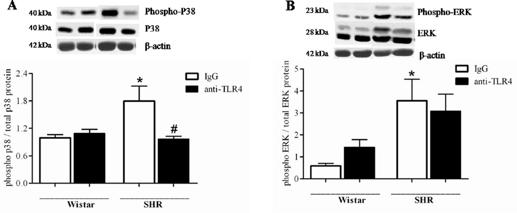
Figure 4. Anti-TLR4 treatment decreases vascular p38 phosphorylation in SHR.
Phosphorylated and total p38 (A) and ERK1/2 (B) protein expression in mesenteric resistance arteries from Wistar and SHR treated with IgG or anti-TLR4. On the top, representative Western blotting images of (A) p38 and (B) ERK1/2. On the bottom, corresponding bar graphs showing the relative expression of p38 and ERK1/2, after normalization to β-actin expression. n=6 in each experimental group. *P < 0.05 compared with Wistar IgG; # P < 0.05 compared with SHR IgG. Statistical test: One-way ANOVA.

