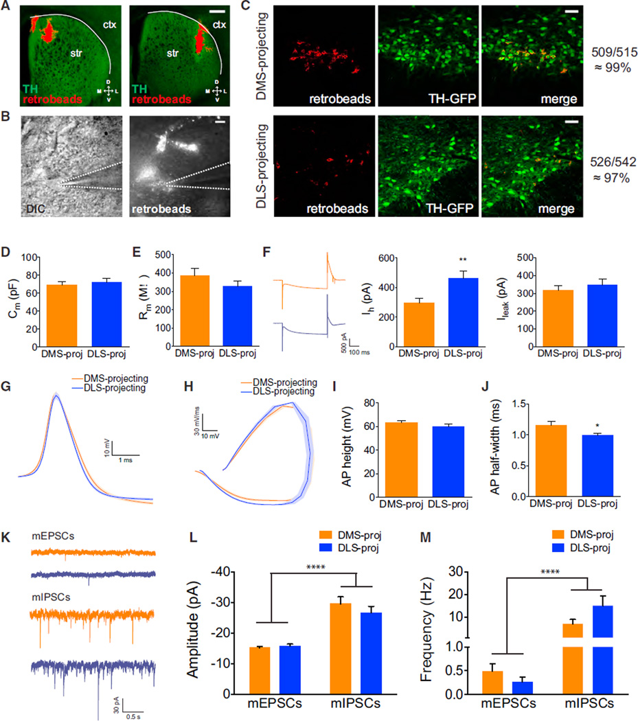
Figure 3. Intrinsic Properties of DMS-Projecting and DLS-Projecting SNc DA Neurons.
(A) Retrobeads (red) injected into the striatum of TH::GFP mice. Ctx, cortex; Str, striatum. Scale bars are 0.5 mm.
(B) DIC and red fluorescent images of a patched retrobead-containing neuron. Dotted lines highlight the position of the recording electrode. Scale bars are 10 µm.
(C) Colocalization of retrobead-containing neurons (red) with TH-GFP-labeled neurons (green) in the SNc. Colocalization rates were ~99% and ~97% in DMS-and DLS-projecting neurons, respectively.
(D) Membrane capacitance (Cm) of DMS- and DLS-projecting SNc DA neurons. Error bars are SEM.
(E) Membrane resistance (Rm) of DMS- and DLS-projecting SNc DA neurons. Error bars are SEM.
(F) Left, example responses of DMS-projecting SNc neurons (orange) and DLS-projecting SNc neurons (blue) to a hyperpolarizing current injection. Right, Ih and Ileak current measurements in response to hyperpolarizing current injection from DMS- and DLS-projecting SNc DA neurons. Error bars are SEM. **p < 0.01.
(G) Average action potential waveforms from DMS- and DLS-projecting SNc DA neurons. Area of light shading is SEM.
(H) Phase plots of average action potential waveforms from DMS- and DLS-projecting SNc DA neurons. Area of light shading is SEM.
(I) Average action potential height from DMS- and DLS-projecting SNc DA neurons. Error bars are SEM.
(J) Average action potential half widths from DMS- and DLS-projecting SNc DA neurons. Error bars are SEM. *p < 0.05.
(K) Example traces of excitatory (top) and inhibitory (bottom) miniature postsynaptic currents from DMS-projecting (orange) and DLS-projecting (blue) SNc DA neurons.
(L) Amplitudes of mEPSCs and mIPSCs recorded from DMS-projecting and DLS-projecting SNc DA neurons. Error bars are SEM. ****p < 0.0001.
(M) Frequencies of mEPSCs and mIPSCs from recorded DMS-projecting and DLS-projecting SNc DA neurons. Error bars are SEM. ****p < 0.0001.
See also Figure S3.

