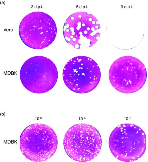
FIG. 2.
Progression of plaque formation with time in Vero and MDBK cells. (a) Plaque formation was assayed after inoculation with 50 PFU of HSV-1 [17] in Vero and MDBK cells as indicated. Monolayers were fixed and stained at 3, 6, and 9 days after infection. (b) Plaque formation was assayed in MDBK cells after inoculation with different dilutions of HSV-1 [17], representing 5,000, 500, and 50 plaques. Monolayers were fixed and stained at 6 days after infection. d.p.i., days postinfection.

