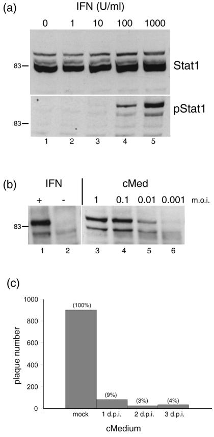
FIG. 5.
Production of IFN in MDBK cells. (a) MDBK cells were treated with medium containing increasing doses of standard uIFN-α (as indicated) and harvested 1 h later. Cell aliquots were then separated by SDS-PAGE and probed by Western blotting for total Stat1 (upper panel) and pStat1 (lower panel). (b) MDBK cells were infected with HSV-1 [17] at the dilutions indicated, medium was harvested 24 h later, and virus was removed by using sterile polyvinylidene difluoride syringe filters and the media were then applied to a fresh uninfected MDBK monolayer. These cells were harvested after 1 h and probed for pStat1 (lanes 3 to 6). Lanes 1 and 2, treatment with uIFN as a control. +, present; −, absent. (c) MDBK cells were mock infected or infected with 7,000 PFU of HSV-1, and conditioned medium was harvested at 1, 2, or 3 days after infection, filtered, and applied to a fresh MDBK monolayer. Twelve hours later, the conditioned medium was removed and the cells were infected with 1,000 PFU of HSV-1 [17]. Plaques were counted 3 days later. Plaque formation after application of the conditioned medium (cMed, cMedium) is indicated as a percentage relative to that of the control medium (taken as 100%). d.p.i., days postinfection.

