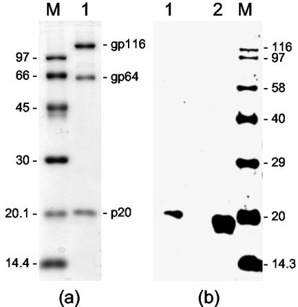
FIG. 4.
(a) Structural proteins (gp116, gp64, and p20) of purified YHV (lane 1) detected by staining with Coomassie blue. (b) Western blot of purified YHV (lane 1) and GAV-infected gill tissues (lane 2) with GAV GST-ORF2 antiserum. Proteins were resolved in a 12% polyacrylamide gel. Lanes M contain protein standards (a) and biotinylated protein standards (Amersham) (b), with molecular masses (in kilodaltons) indicated.

