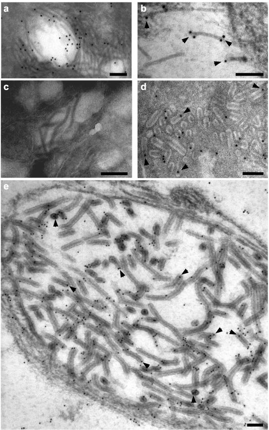
FIG. 5.
Immunoelectron microscopy of ultrathin sections of GAV-infected LO cells using prebleed (c) or KLH-PN1 peptide (a, b, d, and e) antiserum. No gold particles (10 nm in diameter) associated with GAV nucleocapsids with prebleed sera. With the PN1 peptide antiserum, gold particles associated with free striated nucleocapsids (a and b) and nucleocapsids within mature rod-shaped virions (d) and newly formed virions (e) that were observed budding into a membranous endoplasmic vesicle. Good examples of gold particles bound to nucleocapsid ends or to areas where the nucleocapsids had been cross-sectioned laterally are highlighted (arrowheads). Bars represent 100 nm.

