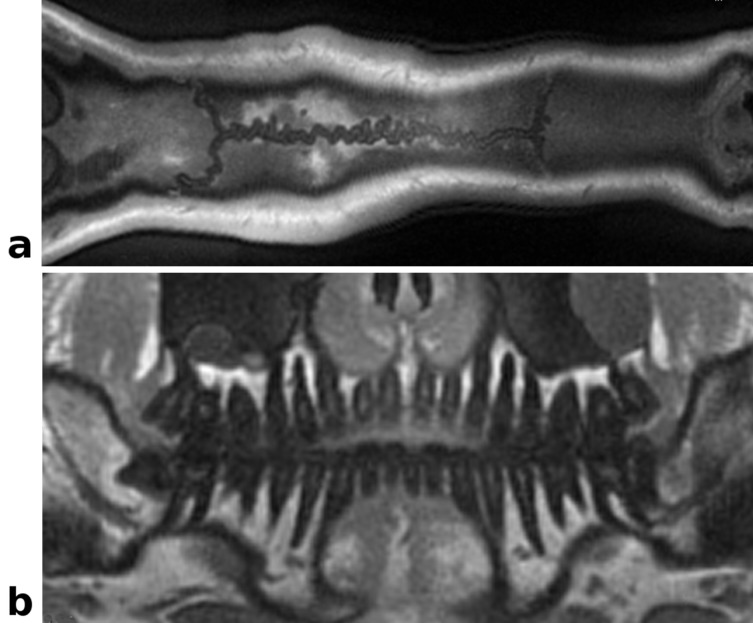
Fig 6. Curved reformatted In vivo head images: Re-sliced UTE images obtained from the same data set as in Fig 6.
(a) Curved reformatted slice displaying the sagittal suture. (b) Curvilinear reformatted view of the denture, mimicking a dental panorama image. The nerve canals, dentin and the jawbone are clearly delineated.

