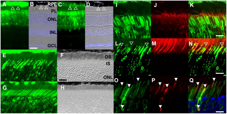
Fig 6. C21orf2 localized to the connecting cilium of the rod and cone photoreceptors.
(A-D) Expression of EGFP driven by CMV-promoter or C21orf2-promoter. When driven by the ubiquitous CMV-promoter, EGFP showed stronger expression in the retinal pigment epithelium (RPE; Open triangle) than in the photoreceptors (A, B). When driven by the C21orf2-promoter, EGFP is expressed more prominently in photoreceptors than in RPE (C, D). (E-H) AAV8-mediated expression of EGFP fusion protein. The C21orf2-EGFP fusion protein was not detected in the outer segments (OS; E, F), while EGFP was present in the OS in the control (G, H). (I-K) C21orf2 localized to the connecting cilium (red; stained with anti-acetylated-tubulin antibodies). (L-N) Association of C21orf2 to the connecting cilium, but not to the surrounding OS structure in cone photoreceptors. C21orf2-EGFP fusion protein remains localized to the cilia (open arrowheads) inside the PNA-positive cone OS (red). (O-Q) Lack of spatial association between C21orf2 and mitochondria. Kusabira Orange-tagged mitochondria (red). RPE, retinal pigment epithelium; PL, photoreceptor layer; ONL, outer nuclear layer; INL, inner nuclear layer; GCL, ganglion cell layer; OS, outer segment; IS inner segment. Scales bars: 50 μm (B), 30 μm (H, K, N) and 15 μm (Q).

