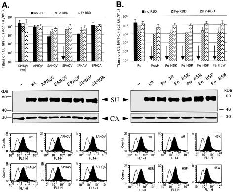
FIG. 2.
Mutagenic analysis of the conserved amino-terminal SPHQV motif of the FeLV-B envelope glycoprotein. (A) Each amino acid of the SPHQV motif was replaced individually by an alanine. (B) The histidine of this motif was deleted (ΔH) or replaced with a basic (K or R) or aromatic amino acid (F or W). The bar graphs show infectivities of the wild-type and mutant viruses in CE-Pit1 cells in the absence or presence of FeLV-B RBD (Fe-RBD) or of the Friend MuLV RBD (Fr-RBD). The titers are averages of four independent experiments ± standard errors of the means. The middle data are Western immunoblots of the wild-type and mutant pseudotyped virus pellets, showing the SU glycoproteins and the viral capsid (CA) proteins. The bottom data show FACS analyses of the binding of the wild-type and mutant envelope glycoproteins onto MDTF/FePit1 cells. The background fluorescence (black lines) was measured when the cells were incubated with media from nontransfected TELCeb6 cells. The assays were done three times. wt, wild type.

