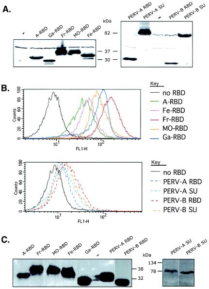
FIG. 4.
Characterization of RBD and SU envelope glycoproteins. (A) 293T/mCAT1 cells that had been stably transfected with RBD and SU expression vectors were used for analysis of the cell-associated envelope glycoproteins by Western immunoblotting as previously described (28, 31, 34) using the anti-RGS(H)4-specifc mouse antibody. (B) Conditioned medium from negative control TE671 cells or cells making RBDs or SUs were incubated with EDTA-detached 293T/mCAT1 cells for 1 h at 37°C before washing the cells and incubating them with the anti-RGS(H)4 antibody. The data show FACS analyses. (C) The conditioned media used for the latter binding assays were analyzed directly by Western immunoblotting with the anti-RGS(H)4 antibody. The RBDs were shed into the culture media in similar amounts, although they bound to the 293T/mCAT1 cells to different extents. Ga, GALV; Fe, FeLV-B; Fr, Friend MuLV; MO, Mo-MuLV; A, amphotropic strain 4070A MuLV.

