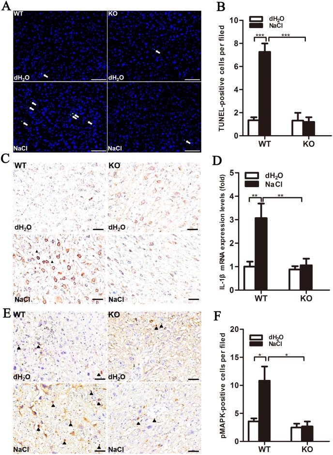
Fig 5. Increased numbers of apoptotic cells in placenta labyrinths of high salt-treated WT mice through IL-1β signaling pathway activation.
(A) TUNEL-positive cells (green, arrows) were observed in placenta. DAPI (blue) was used for nuclear staining. Scale bar = 100 μm. (B) DAPI/TUNEL double-positive cells in placenta labyrinth were quantified in 10–15 fields/placenta and averaged. n = 4. Immunohistochemical imaging of IL-1β (C) and Il1b mRNA expression (D), detected by real-time qPCR (n = 5/group). (E) Immunohistochemical imaging of phosphorylated p38-MAPK (arrowhead show positive signals, hematoxylin co-stained with nucleus). Scale bar = 500 μm. (F) Positive cells were quantified in 10–15 fields/placenta and averaged (n = 4/group).

