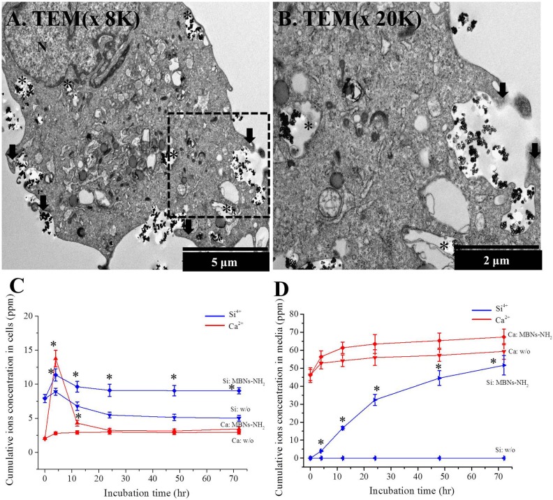
Fig 4.
(a, b) Macropinocytosis of MBNs–NH2 into rDPSCs after 4 hours of incubation by TEM images and Ca and Si ion concentrations from total cells (c) or cultured odontogenic supplemental media (OM) (d) by ICP-AES. Low-magnification (a, ×8,000) and high-magnification (b, ×20,000, black dot box from (a)) TEM images of rDPSCs after treatment with MBNs–NH2 (50 μg/mL of 105 cells) for 4 hours. Macropinocytosis with membrane ruffles was only observed without clathrin or caveolae-coated membrane or vesicle. Black arrow shows membrane ruffles, a typical characteristic of macropinocytosis; black asterisks indicate the intracellular distribution of MBNs–NH2 in endosomes. Significant increases in Si ion concentration from total cells were detected at 12, 24, 48, and 72 hours in the MBNs–NH2–treated rDPSCs compared to control (without [w/o] treatment), while Ca ion concentrations were significantly detected at 4 and 12 h of incubation (p<0.05). According to the preliminary results for cell numbers at 4, 12, 24, 48, or 72 hours of incubation under OM with or without MBNs–NH2, there was no significant difference. * Statistically significant difference in MBNs–NH2–treated group compared with the untreated group (p<0.05).

