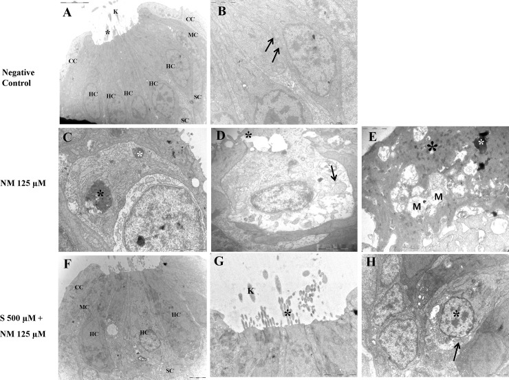
Fig 6. Transmission electron microscopy (TEM).
TEM of Normal control (A and B); TEM of zebrafish treated with 125 μM neomycin only (C-E); TEM of zebrafish treated with 125 μM neomycin and 500 μM sodium selenite (F-H). The sterocilia (black asterisk) and the kinocilium (K) from each hair cell are clearly visible (× 8K) (A). A normal-sized mitochondria (arrow, × 25K) (B). The hair cells were severely damaged and showed a condensed nuclei (black asterisk) and pyknotic nuclei (white asterisk) (× 20K) (C). The collapse of the apical surface of neuromasts was typically evident. A hair cell showed extrusion of cytoplasm (black asterisk) and the swollen mitochondria (arrow) (× 20K) (D).Severely damaged hair cells show a severely degenerating cytoplasm (black asterisk), a fragmenting condensed nucleus (white asterisk), and multiple swollen mitochondria (M) (× 30K) (E).When 500 μM sodium selenite was applied, structure of neruomasts were nearly complete protected. Nuclear damage such as condensed cytoplasm and pyknotic nuclei were not showed. (× 5 K) (F). The structure of the stereocilia (black asterisk) and the kinocilium(K) are preserved (× 20 K) (G). The normal-sized nucleus (black asterisk) and mitochondria (arrow) is shown (× 20 K) (H). NM, neomycin; S, sodium selenite; HC, hair cell; SC, supporting cell; CC, crescent cell; MC, mantle cell; BM, basement membrane. Scale bars (at the top or the bottom of each figure, one space) = 5μm (A); 2μm (B); 2μm(C); 1μm (D); 1 μm (E); 2μm (F); 2μm (G); 2μm (H). NM, neomycin; S, sodium selenite. Images were obtained in three 5-dpf zebrafish for each group.

