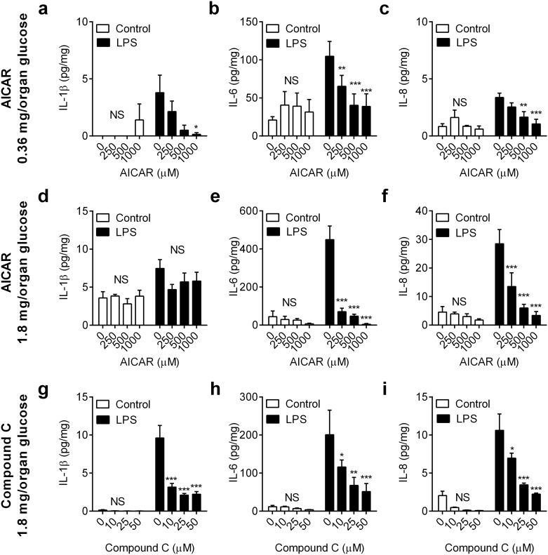
Fig 4. Manipulation of AMPK activity regulates inflammation in endometrial tissue.
Ex vivo organ cultures of endometrium were cultured for 24 h in medium containing 0.36 mg/organ glucose (a-c) or 1.8 mg/organ glucose (d-f) with vehicle (0) or AICAR (250, 500, 1000 μM) to activate AMPK, or (g-i) in medium containing 1.8 mg/organ glucose with vehicle (0) or Compound C (10, 25, 50 μM) to inhibit AMPK. Media was then aspirated and replenished with fresh medium containing a corresponding concentration of glucose and AICAR or Compound C, and challenged with control vehicle or 100 ng/ml LPS for a further 24 h. At the end of the experiment, organ weights were recorded, and the accumulation of IL-1β (a, d, g), IL-6 (b, e, h) and IL-8 (c, f, i) was measured in supernatants. Data are presented as mean concentration per mg tissue + SEM from 4 independent experiments, and analyzed by ANOVA using Dunnett’s multiple comparisons test to compare with vehicle (0), within each treatment group; * P < 0.05, ** P < 0.01, *** P < 0.001, NS = ANOVA not significant.

