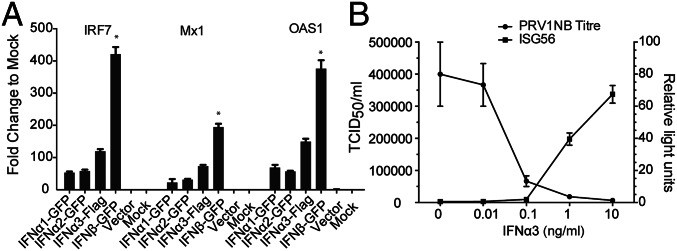
Fig. 4.
Bat IFN-α proteins are functional. (A) Bat IFN-α proteins induce an ISG response. Bat IFN-α and IFN-β protein containing supernatant produced in HEK293T cells were used to treat PaKiT03 cells. Six hours later, cells were collected for qRT-PCR detection of mRNA expression of IRF7, Mx1, and OAS1. Supernatant from empty vector-transfected or mock-transfected HEK293T cells were used as controls. Data show fold changes compared with mock and represent the mean and SD from two experiments. The difference in ISG induction in response to IFN-β was calculated in comparison with ISG induction by each of the three individual IFN-α proteins (*P < 0.05). (B) Bat IFN-α3 blocked PRV1NB replication in a dose-dependent manner. IFN-α3 protein was added at varying doses to PaKiT03 cells before PRV1NB infection, and 50% tissue culture infective dose (TCID50) assays were performed. In a parallel experiment, PaKiT03 cells were transfected with bat ISG56 promoter reporter plasmid, and luciferase activity was analyzed after treatment with varying doses of IFN-α3. Fold activation was determined by dividing the relative light units of each experimental sample by the relative light units of media alone. Data represent the mean and SE of triplicate experiments.

