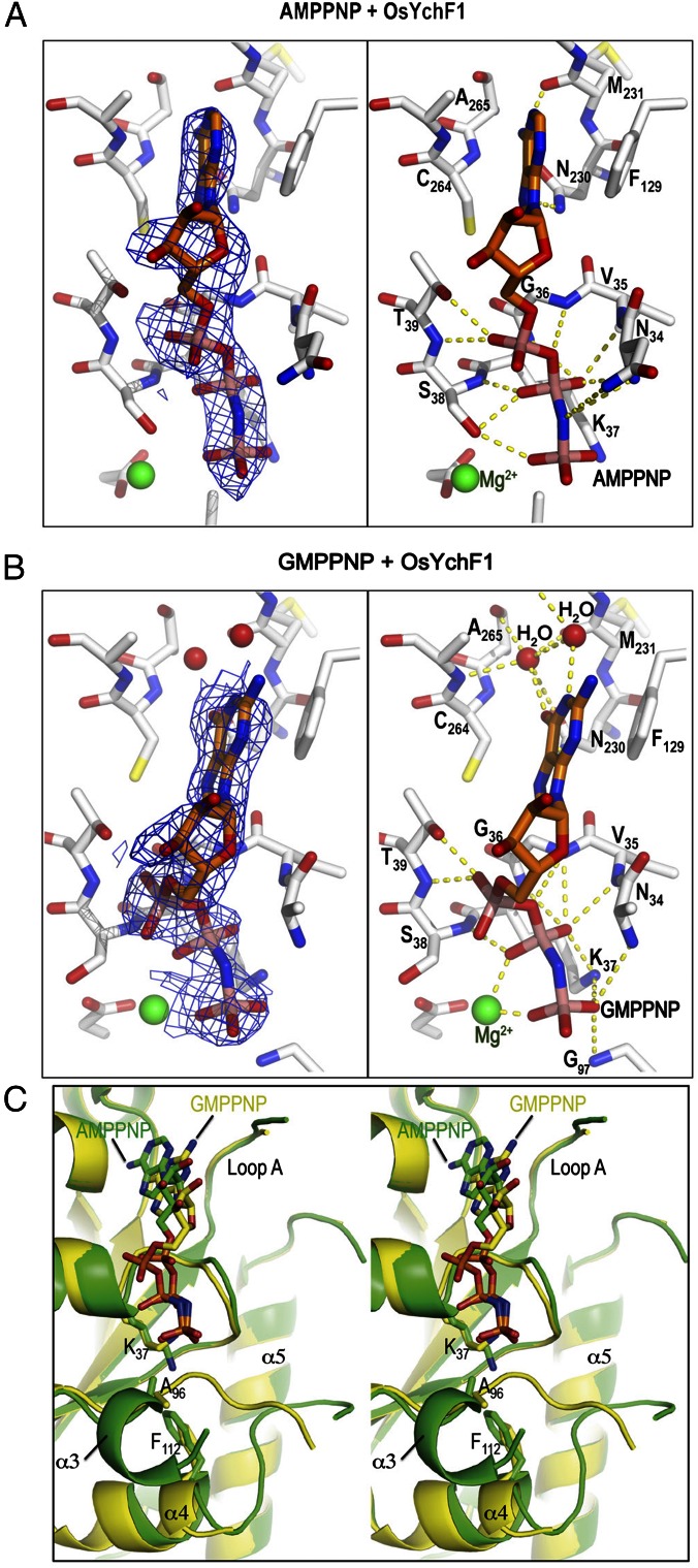
Fig. 2.
Structures of OsYchF1 cocrystallized with AMPPNP and GMPPNP. (A and B) Structural diagrams of OsYchF1 cocrystallized with AMPPNP (A) and GMPPNP (B). The “omit” electronic density of nucleotides are contoured at 1σ at Left, whereas hydrogen bonds between the protein and the nucleotides are indicated as yellow dotted lines at Right. (C) A stereo diagram showing the structural comparison of OsYchF1 in complex with AMPPNP (PDB ID code: 5EE3, green) and GMPPNP (PDB ID code: 5EE9, yellow). The guanine base is bound to the binding pocket ∼2 Å shallower than the adenine base. Binding of GMPPNP induces the unfolding of the helix α3 so that the backbone amide of Gly-97 of the G3/switch II region can form a hydrogen bond with the γ-phosphate of GMPPNP.

