Fig. 1.
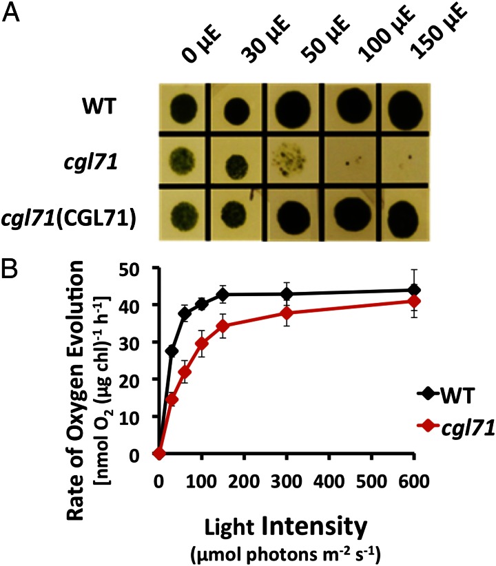
Growth and O2 evolution of the cgl71 mutant. (A) WT, cgl71, and cgl71(CGL71) rescued cells were grown at 30 µmol photons⋅m−2⋅s−1 (µE) in liquid TAP medium and then were washed and spotted onto solid TAP medium and allowed to grow for 5 d at various light intensities, as indicated at the top of the image. (B) Rate of O2 evolution of WT and cgl71 mutant cells after growth at 30 µE. Cells were pelleted by centrifugation (3,200 × g) for 10 min, resuspended in 50 mM Hepes (pH 7.5) containing 5 mM NaHCO3, and shaken in the dark for 30 min. During the assay, samples were illuminated at increasing light intensities, as indicated on the x axis, for 2 min each, followed by a 2-min dark incubation. The rate of O2 evolution at each light intensity was corrected for the rate of dark respiration. Each point represents the mean of three biological replicates.

