FIGURE 5:
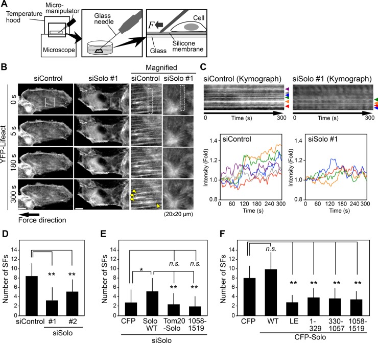
Solo is required for tensional force–induced stress fiber formation. (A) Methods of tensional force application and time-lapse observation. (B) Solo knockdown suppresses force-induced stress fiber formation. MDCK/YFP-Lifeact cells were plated on a silicone membrane, transfected with control or Solo-targeting siRNAs, and cultured for 48 h before tensional force was applied. Time-lapse fluorescence images of YFP-Lifeact near the ventral surface of the cell were obtained by confocal microscopy. Right, magnified images of the white boxes in the images on the left. Yellow arrowheads indicate stress fibers that were strengthened or emerged after force application. Scale bars, 20 μm. See also Supplemental Movie S1. (C) Kymographs of the dashed boxes in B. Bottom, changes in fluorescence intensity of each of stress fiber indicated by arrowheads in the kymographs. (D–F) Quantitative analysis of the effects of knockdown of Solo (D), knockdown of Solo followed by expression of Solo-WT or its mutants (E), or expression of Solo or its mutants (F) on force-induced stress fiber formation. The number of stress fibers that were newly generated or reinforced was counted. See also Supplemental Figures S3C and S4B and Supplemental Movies S1–S3. The data shown represent the mean ± SD of 16–19 cells/experiment. *p < 0.05 and **p < 0.01; n.s., not significant (one-way ANOVA followed by Dunnett’s test or Tukey’s test).

