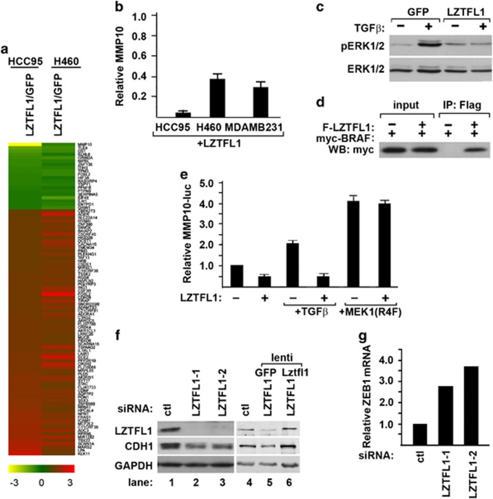
Figure 4.
LZTFL1 inhibits tumor cell EMT. (a) Heat map of genes differentially expressed (greater than or equal to twofold) in LZTFL1-expressing HCC95 and H460 cells compared with GFP-expressing cells. (b) Transcripts of MMP10 in LZTFL1-GFP expressing HCC95, H460 and MDA-MB-231 cells measured by qRT–PCR. Transcripts were expressed relative to that in control GFP-expressing cells. (c) H2347 cells were transduced with lentiviral vectors expressing GFP or LZTFL1 and treated with or without TGFβ. Cells were harvested 30 min later for western blotting (WB) analysis with pERK1/2 and ERK1/2 antibodies. (d) myc-BRAF was transfected without or with Flag(f)-LZTFL1 into 293 T cells. Cell lysates were immunoprecipitated (IP) with anti-flag antibody and western blotted with anti-myc antibody. In all, 10% of input was loaded on the gel. (e) MMP10-luciferase activities from H2347 cells transfected with or without LZTFL1, stimulated with or without TGFβ or co-transfected with or without MEK1(R4F). (f, g) Control siRNA (ctl) or LZTFL1-specific siRNAs (LZTFL1-1 and -2) were transfected into H2347 cells. Cell lysates were harvested 72 h later for WB (f) and qRT–PCR analysis (g). For rescuing experiment in panel (f), lentiviruses expressing either GFP or mouse-Lztfl1 were transduced into LZTFL1-siRNA-transfected cells 24 h after transfection.

