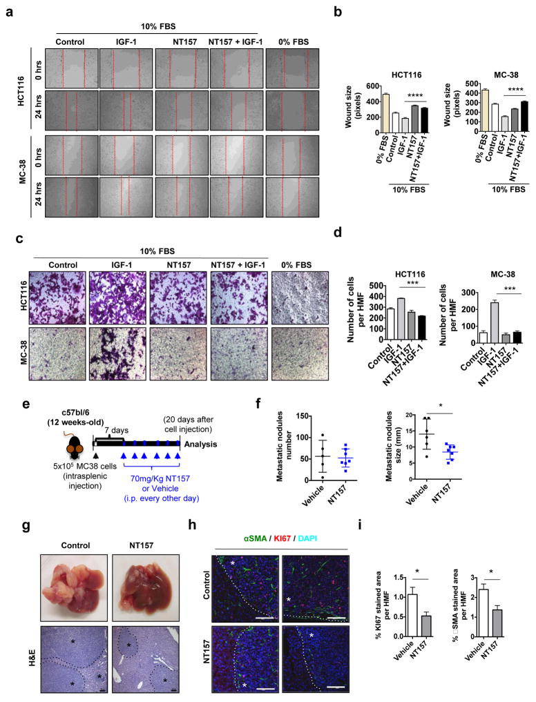
Figure 4. NT157 treatment decreases the metastatic activity of CRC cells.
a, b. HCT116 and MC-38 cells were seeded and grown to confluency. The cell monolayers were scratched and the cells were left to grow in the presence of IGF-1 (100 ng/ml) with or without NT157 (3 μM). Images were taken at 0 and 24 hrs, and wound size was determined after 24 hrs using Axo Vision software (distance between the edges in pixels). Three different measurements were recorded from 7–10 fields per condition n=2 independent experiments. **** p<0.001 vs. IGF-1 stimulated cells c, d. HCT116 and MC-38 cell migration was determined using Boyden chambers. Briefly, 2×10⁵ viable cells were seeded on the top compartment and left to attach for 2 hrs, afterwards cells were allowed to migrate in DMEM supplemented with 10% FBS in the presence or absence of NT157 and after adding IGF-1 to the lower compartment as a chemotactic agent. After 6 hrs, transmigrated cells were stained using crystal violet, photographed and the number of cells per 6 HMF was calculated for each condition. Results are averages ± s.e.m. *** p<0.005. e. MC-38 cells were injected i.s. into C57BL/6 mice. Seven days later the mice were treated with 70 mg/Kg NT157 or vehicle as indicated and sacrificed 20 days after cell inoculation. f. Metastatic lesions in liver were enumerated and measured. Results are means ± s.e.m. (n=5–7 mice per group). * p<0.05. g. Representative images of livers from above mice and H&E staining of liver sections (scale bars: 100 μm) * Indicates tumor tissue h. Paraffin embedded liver sections were IF analyzed after staining with indicated antibodies (scale bars: 100 μm) * Indicates tumor tissue. i. Positively stained areas per each HMF (high magnification field) were quantitated using ImageJ Software. Results are averages ± s.e.m. * p<0.05. (n=7 HMF per group).

