
Figure 2. Exercise training and EPO counteract oxidative fiber atrophy and glycolytic shift stimulating PGC-1α expression.
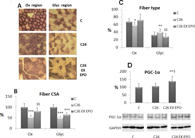
A. SDH (succinate dehydrogenase) staining in cross sections of tibialis muscle from control (C), C26-bearing (C26) and C26 exercised EPO-treated (EX EPO) mice (2 weeks of exercise). The two micrographs for each group represent two regions with distinct frequency of oxidative (Ox) and glycolytic (Glyc) fibers. B. Morphometric analysis of myofiber CSA (cross-sectional area) performed on SDH stained sections. Data (mean±SD) are expressed as percentages of controls. C. Quantification of fiber type frequency in the tibialis muscle. Data (mean±SD) are expressed as relative percentages. D. PGC-1α nuclear protein expression in the tibialis muscles. Densitometric quantifications were normalized according to GAPDH levels. Data (mean±SD) are expressed as percentages of controls. Significance of the differences: *p < 0,05 vs C, **p < 0,01 vs C, ***p < 0,001 vs C, $p < 0,05 vs C26, $$p < 0,01 vs C26.