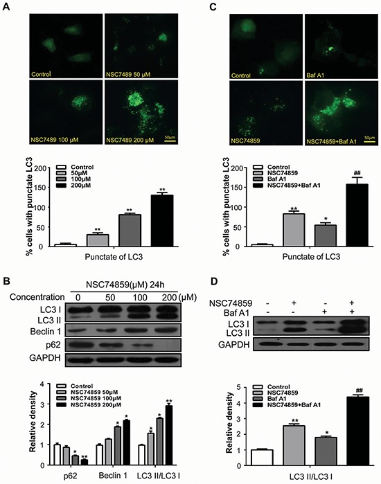
Figure 2. Targeting STAT3 by NSC74859 induced autophagy in HNSCC cells.

A. CAL27 cells transfected with GFP-LC3 plasmid were treated with different concentration of NSC74859 for 24 h. The formation of GFP-LC3 puncta were examined using immunofluorescence and quantified. Scale bar 50 μm; **P < 0.01; B. CAL27 cells were treated with different concentration of NSC74859 for 24 h, then detected autophagy-associate protein LC3I/II, p62, and Beclin1 by western blot analysis; Densitometric values were quantified using the Image J software, and the data were presented as means ± SEM of three independent experiments. *P < 0.05, **P < 0.01; C. CAL27 cells were treated with 100 μM of NSC74859 in the absence or presence of 20 nM Bafilomycin A1 for 24 h, then formation of GFP-LC3 puncta were examined using immunofluorescence and quantified, *P < 0.05, **P < 0.01 versus the control group, One-way ANOVA with post-Dunett analysis was used by GraphPad Prism5; ##P < 0.01 versus the NCS74859 (100 μM) group, One-way ANOVA with post-Tukey analysis was used by GraphPad Prism5; D. CAL27 cells were treated with 100 μM of NSC74859 in the absence or presence of 20 nM Bafilomycin A1 for 24 h, then the expression of LC3II was quantified by normalization of their densitometry to GAPDH; Densitometric values were quantified using the Image J software, and the data were presented as means ± SEM of three independent experiments. *P < 0.05, **P < 0.01 versus the control group, One-way ANOVA with post-Dunett analysis was used by GraphPad Prism5; ##P < 0.01 versus the NCS74859 (100 μM) group, One-way ANOVA with post-Tukey analysis was used by GraphPad Prism5.
