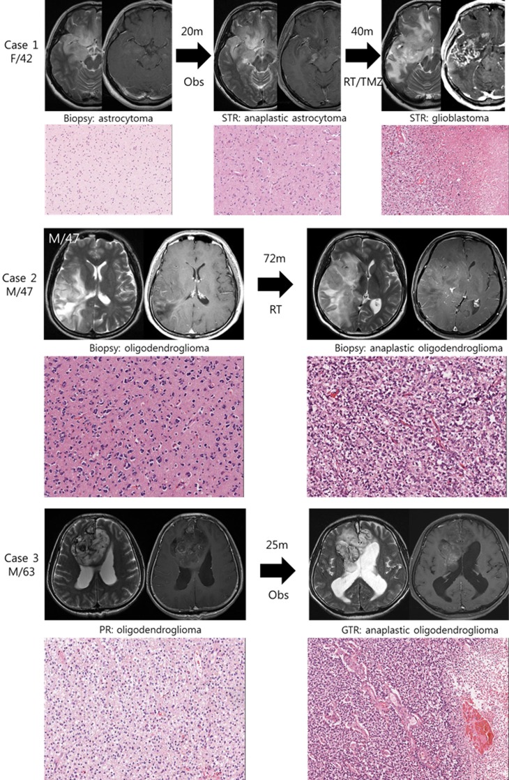
Figure 1. Illustrative history of patients.
Case 1. A 42-year-old female patient who was initially diagnosed with diffuse astrocytoma in right fronto-temporal area after stereotactic biopsy. At the option of the patient, she was monitored without any treatment and was in stable state for 20 months. However, enhancing area in magnetic resonance images (MRI) was developed in temporal lobe lesion, and subtotal resection was performed. The diagnosis was anaplastic astrocytoma, and she was treated with radiotherapy followed by 6 cycles of temozolomide chemotherapy. Forty months after the second surgery, progression of the disease was observed and the diagnosis of glioblastoma was confirmed after the third surgery. A pair of diffuse astrocytoma and glioblastoma samples were used in this study. Case 2. A 47-year-old male patient with right temporo-parietal mass was diagnosed with oligodendroglioma after stereotactic biopsy and was treated with standard radiotherapy alone. The mass was aggravated to frontal area after 72 months of stable period, and the anaplastic oligodendroglioma was confirmed after stereotactic biopsy. Case 3. A 63-year-old male patient with heterogeneous bifrontal mass lesion was diagnosed with oligodendroglioma after partial resection, and the remaining mass was observed over period. After 25 months of stable period, the disease started to progress and the second surgery was done. The histological diagnosis was confirmed to be anaplastic oligodendroglioma after gross total resection.

