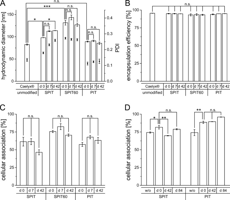
Figure 3.
(A) Hydrodynamic diameter (primary y-axis) and PDI (secondary y-axis) of Caelyx® determined by photon-correlation-spectroscopy before and after surface modification via SPIT, SPIT60 and PIT (d 0), as well as after 1 week (d 7) and 6 weeks (d 42) of storage at 4°C (n = 3 ± SEM), (B) Encapsulation efficiency of Dox in Caelyx® before and after surface modification via SPIT, SPIT60 and PIT (d 0), as well as after 1 week (d 7) and 6 weeks (d 42) of storage at 4°C (n = 3 ±SEM), (C) Cellular association of Rh-PE labelled HSPC/Chol/DSPE-mPEG liposomes dispersed in SH buffer modified via SPIT, SPIT60 or PIT using an anti GD2 ab with Kelly cells at 37°C, immediately after production (d 0) and after 1 and 6 weeks of storage (d 7 and d 42), (n = 2 ± SEM), (D) Cellular association of Rh-PE labelled SPC/Chol liposomes dispersed in HBS modified via SPIT or PIT using an anti GD2 ab with Kelly cells at 37°C, without (w/o) and with lyophilisation of ab-anchor conjugate (d 0) and its storage over 6 (d 42) and 12 (d 84) weeks at 4°C (n = 3 ± SEM).

