
Figure 2. GA increases LC3-II expression and GFP-LC3 punta formation in A549 and NCI-H1299 cells.
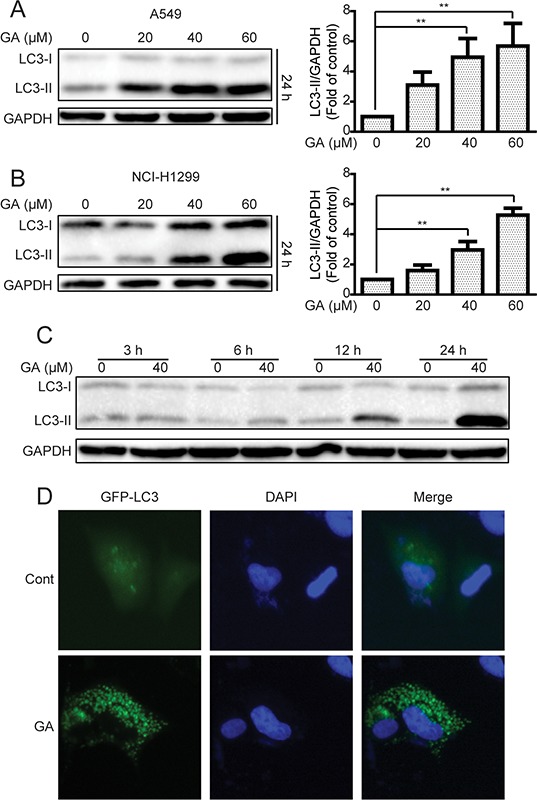
A–B. A549 and NCI-H1299 cells were treated with various concentrations of GA for 24 h, and cell extracts were analyzed to determine changes in protein expression by Western blot analysis. *P < 0.05 and **P < 0.01. C. A549 cells were treated with GA (40 μM) for the indicated time. Western blot was used to detect protein expression. D. A549 cells were transiently transfected with the GFP-LC3 plasmid with or without GA (40 μM) for 24 h. GFP-LC3 punta formation was determined using In Cell Analyzer 2000, and typical images were presented.