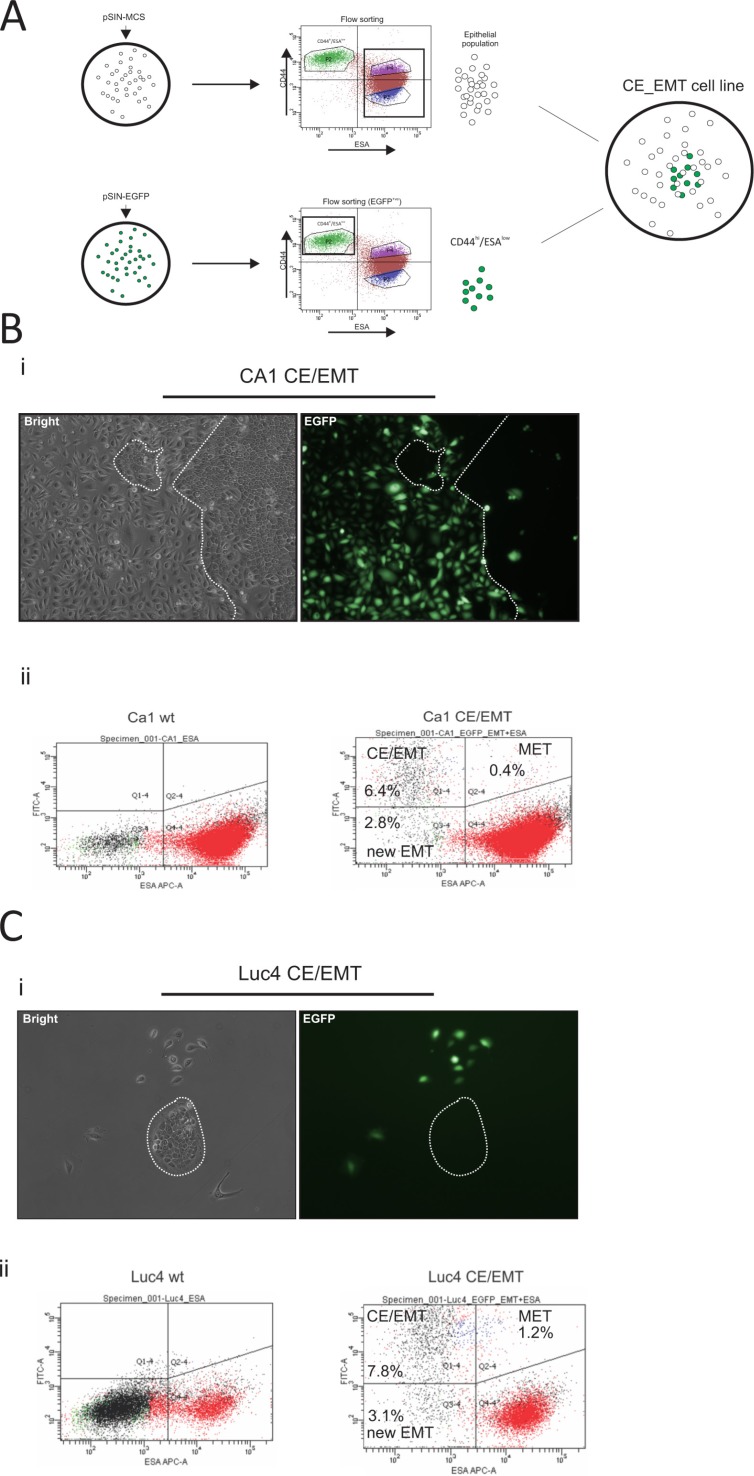
Figure 4. Generation and characterisation of EMT-CE cell lines.
CA1 and Luc4 parental cell lines were transduced with either pSIN-MCS or pSIN-EGFP retroviral vectors (A). pSIN-MCS transduced cells were completely depleted of CD44hi/ESAlow population, while the pSIN-EGFP transduced cell lines were flow sorted and only CD44hi/ESAlow were isolated. Both types were mixed at a ratio which mimicked that of the parental wild-type cell line. (B, C) Cells were later grown for up to 14 days. By 7 days of culture, the very distinct phenotype of mesenchyme-like (CD44hi/ESAlow) cells was almost always accompanied by clear expression of EGFP marker protein (Bi, Ci) whereas epithelial cells did not display expression of EGFP. By 14 days of culture, a minority of EGFP(+) cells with epithelial morphology as well as a significant number of EGFP(−) cells with a mesenchymal phenotype became evident. After prolonged culture we were able to visualise the dynamics of epithelial to mesenchyme transition (EMT) by tracing the EGFP(−) CD44hi/ESAlow cells (Bii, Cii) as well as of mesenchyme to epithelial transition (MET) by tracing the EGFP(+) CD44hi/ESAhi cells. The MET rate appeared to be higher for Luc4 cells compared to CA1 cells, while the rate of EMT seemed to be similar to both types of cells. Continuous monitoring of both reconstituted cell lines showed that a part of the original CD44hi/ESAlow population remained permanently at a mesenchymal state while the remaining population was recycled within the parental cell line.

