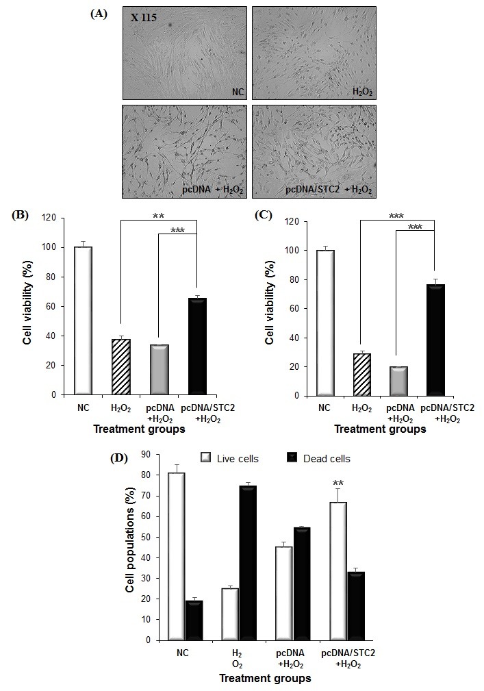
Fig. 2. Increased cell proliferative activity and enhancement of live/dead cell populations by STC2 expressed. At 48 hrs post-STC2 delivery, H2O2 was treated to cells for 3 hrs and then cell morphology was observed (A) and MTT assay were performed (B). Another groups were assessed at 4 days post-media change of H2O2 treatment (C). Increased cell proliferation and viability were observed in the cells transfected with STC2 plasmid. Data represent the means and standard errors of triplicate experiments. **P < 0.02 and ***P < 0.01 for comparison of pcDNA/STC2+H2O2 groups against H2O2 or pcDNA+H2O2 groups, respectively. Also, cell viability of ADSCs was observed by Arthur image-based cytometer in cells stained with PI solution after the treatment of STC2 and H2O2 (D). Dead cells were stained by PI as red color. **P < 0.02 versus H2O2 groups.