Figure 1.
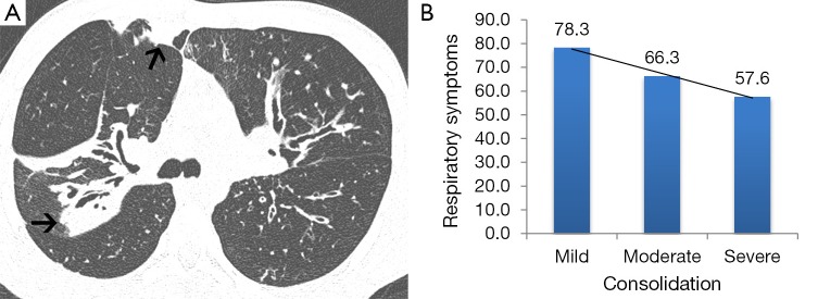
A 15-year-old male outpatient with moderate CF lung disease. (A) Chest CT image shows multifocal consolidation (arrows) in the right lung. (B) Bar graph of Respiratory Symptoms plotted against consolidation on chest CT for all outpatients (R=0.38, P<0.001). This patient scored ‘severe’ for consolidation and scored 44 (poor) on the Respiratory Symptom domain of the CFT-R.

