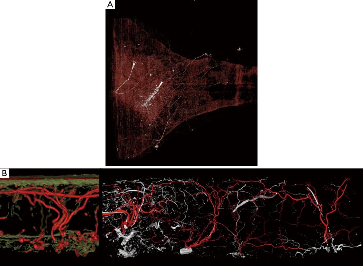
Figure 3.
Examples of human cadaveric vascular injection studies to study the vascular anatomy of a DIEP perforasome using computed tomography (CT) and micro-CT imaging modalities in experimental studies. (A) Human cadaveric injection study of right hemi-abdominal skin flap raised to mid-axial line and ultra-high resolution CT with 0.3 mm slice thickness following injection of a single dominant deep inferior epigastric artery perforator with iodinated contrast media; (B) micro-CT imaging to show high detailing of the relationship of the perforator with the subdermal plexus and recurrent flow to adjacent perforators through direct and indirect linking vessels in the subcutaneous tissue.

