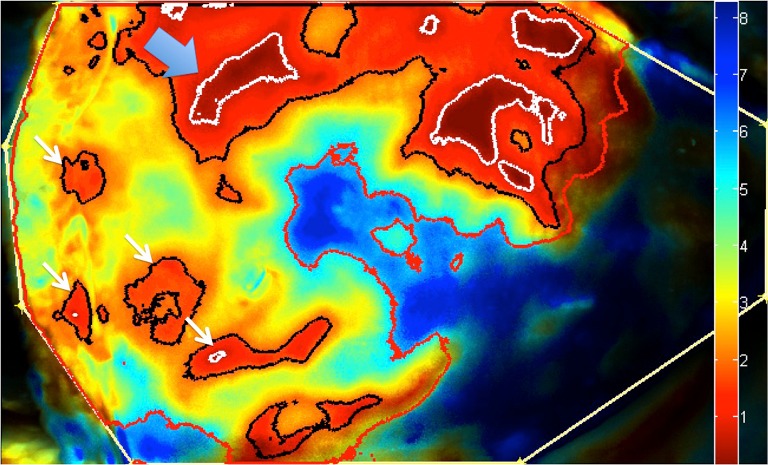
Figure 5.

Laser-assisted indocyanine green fluorescence angiography (LA-ICG) with applied timing color map representing the average ingress of indocyanine green detected across a left hemi- deep inferior epigastric artery perforators (DIEP) flap surface during a 2-minute recording. Flap was harvested on a single dominant perforator (blue arrow), however the timing map may demonstrate the potential location of adjacent perforators (white arrows), receiving blood flow through direct and indirect communicating vessels within the flap.
