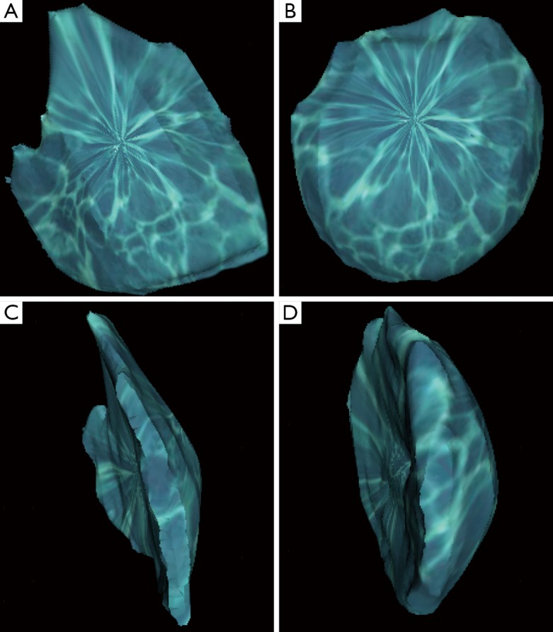
Figure 1.

Volume-rendered 3D reconstructed images of breasts segmented from a computer tomographic (CT) scan using Osirix software (Pixmeo, Geneva, Switzerland), from which volume differential can be calculated. (A) Anterior view of the right breast; (B) anterior view of the left breast; (C) lateral view of the right breast; (D) lateral view of the left breast. Reproduced with permission from Chae et al. (76).
