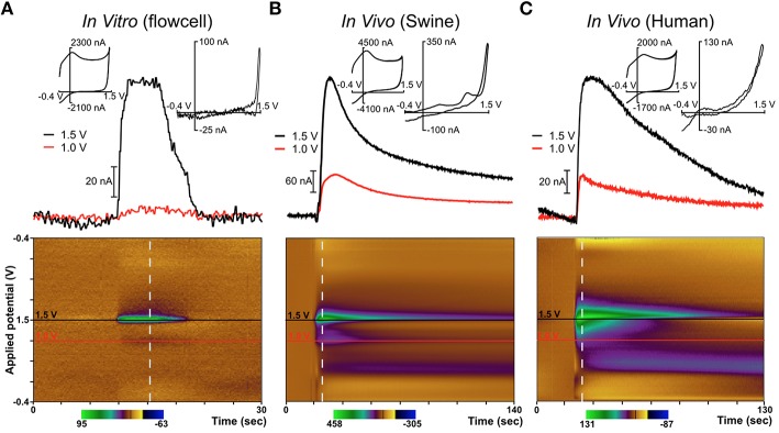
Figure 4.
Diamond electrode performance in vitro and in vivo. Panel (A) depicts a diamond electrode's response to identical in vitro conditions. Panel (B) depicts the same diamond electrode in the ventral lateral thalamus of an anesthetized pig, where an adenosine-like signature secondary to mechanical stimulation was observed. Panel (C) depicts the same electrode in the subthalamic nucleus of an awake human undergoing DBS lead-placement surgery for essential tremor (prior to the DBS lead placement). In panels (B,C), the signature is consistent with adenosine based on the oxidation peaks near the switching potential at 1.5 V. All “diamond” data was recorded with the same specific electrode (although not in the order shown—the human data were always gathered first).

