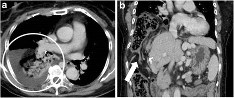
Fig. 2.

a The transverse colon is inserted in the right pleural cavity through the diaphragmatic hernia. The hernia is 10 cm in diameter. Pleural effusion is detected in the right chest cavity (white circle). b The massive transverse colon is intruded into the right chest cavity. Severe stricture can be observed (white arrow)
