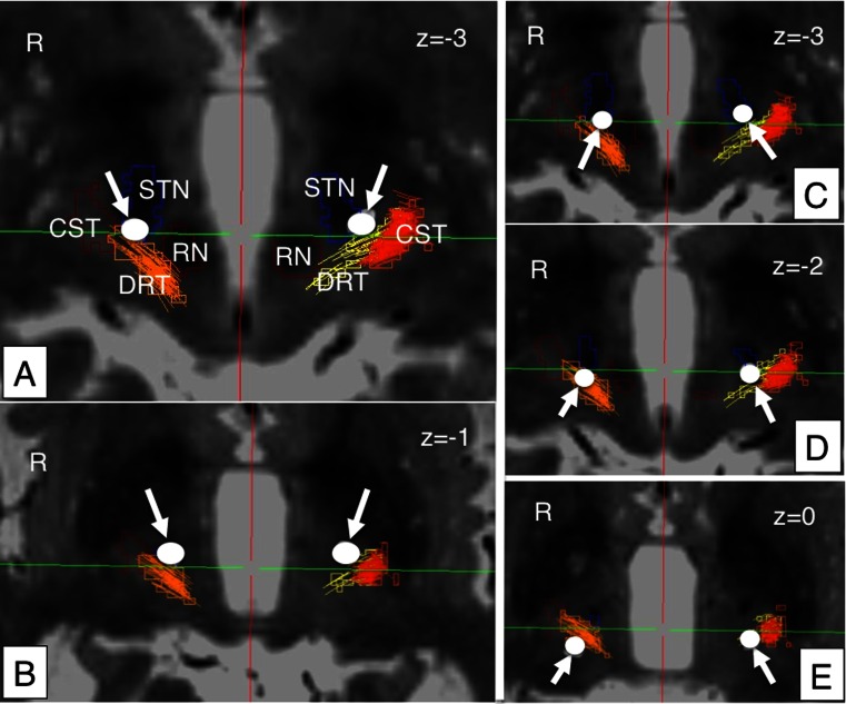
Fig. 3.
Case no. 1, continued. Left column (a, b): Initial implantation attempt without sufficient reduction of upper extremity tremor. DBS electrodes (white arrows) barely touch the DRT, bilaterally. Right column (c, d): Newly proposed parietal approach. DBS electrodes traverse the DRT, bilaterally. ACPC parallel axial views. Z indicates vertical coordinate. Negative z indicates millimeters below ACPC place. DRT dentato-rubro-thalamic tract, CST cortico-spinal tract, RN red nucleus, STN subthalamic nucleus (blue); white arrows (circles) indicate deep brain stimulation electrode contacts

