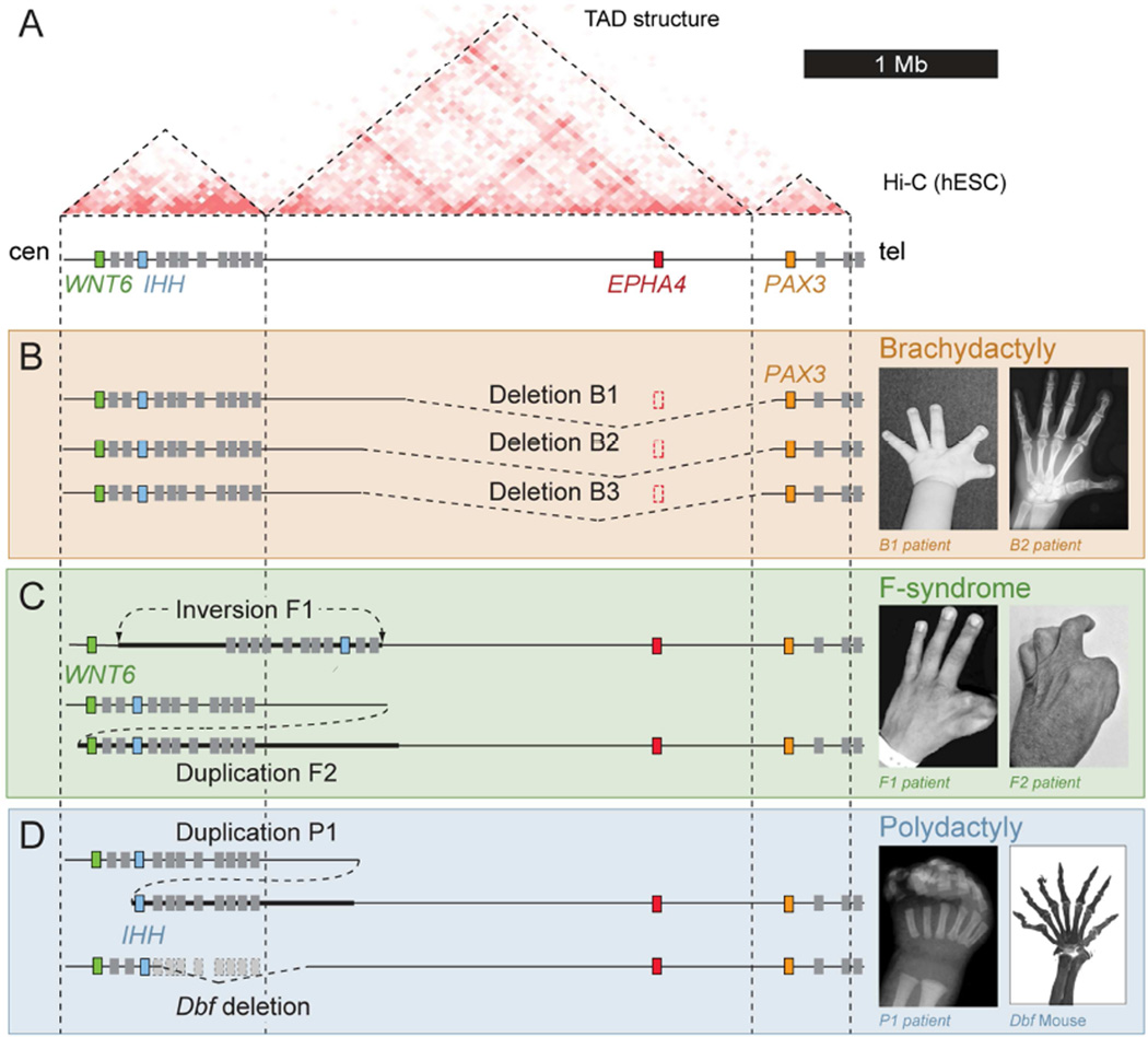
Figure 1. Different limb pathogenic structural variations in human and mouse map to the EPHA4 TAD.
(A) Hi-C profile around the EPHA4 locus in human ES cells (Dixon et al., 2012). Dashed lines indicate the EPHA4 TAD and boundaries. Cen, centromeric; tel, telomeric. (B–D) Schematic of structural variants (left) and associated phenotypes (right). (B) Brachydactyly-associated deletions in families B1, B2 and B3. Note thumb and index finger shortening with partial webbing in a child (B1 patient) and adult (B2 patient). (C) F-syndrome-associated inversion in family F1 and duplication in family F2. Note similar phenotypes of index/thumb syndactyly. (D) Polydactyly-associated duplication (P1) and deletion in the doublefoot (Dbf) mouse mutant. Radiograph of patient hand and skeletal preparation of Dbf/+ mouse show similar 7-digit polydactyly. See also Figure S1.

