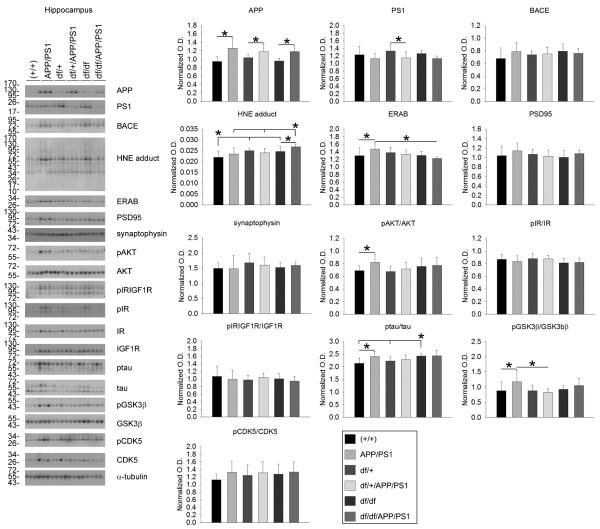
Fig. 4.
df/df/APP/PS1 mice had increased HNE-protein adducts and decreased ERAB in the hippocampus. Hippocampi were collected from wild type (+/+), APP/PS1, dwarf het (df/+), dwarf het APP/PS1 (df/+/APP/PS1), dwarf (df/df), and dwarf APP/PS1 (df/df/APP/PS1) mice. A) The tissues were lysed, resolved by 10% SDS-PAGE and western blotted using anti-APP, PS1, BACE, HNE protein adduct, ERAB, PSD95, synaptophysin, phospho-Akt, Akt (loading control), phospho-IR/IGF1R, phospho-IR, IR (loading control), IGF1R (loading control), ptau, tau (loading control), pGSK3β, GSK3β (loading control), pCDK5, CDK5 (loading control) or α-tubulin (loading control) antibodies. Antibody binding was visualized by chemiluminescence. Blots from 3 animals per group are shown. Optical densities of the western blotted proteins were normalized against their respective loading control and averaged (+/− SD) from 8-13 animals per each condition and graphed, *p<0.05.

