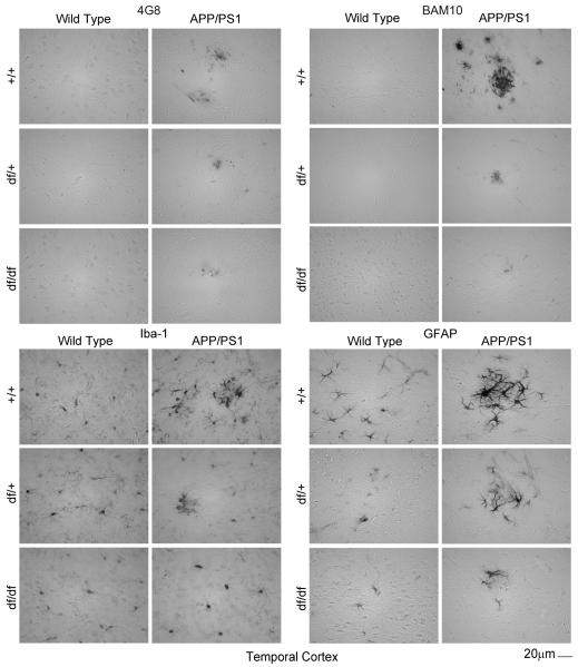
Fig. 7.
df/df/APP/PS1 mice had attenuated Aβ plaque deposition and gliosis in the temporal cortex. Brains were collected from wild type (+/+), APP/PS1, dwarf het (df/+), dwarf het APP/PS1 (df/+/APP/PS1), dwarf (df/df), and dwarf APP/PS1 (df/df/APP/PS1) mice, fixed in 4% paraformaldehyde, serially sectioned, and immunostained. Tissue sections were immunostained using anti-Aβ 4G8 or BAM-10, anti-Iba-1 and anti-GFAP antibodies and antibody binding was visualized using Vector VIP as the chromogen. Representative temporal cortex images from 8-13 mice in each group are shown.

