Figure 4.

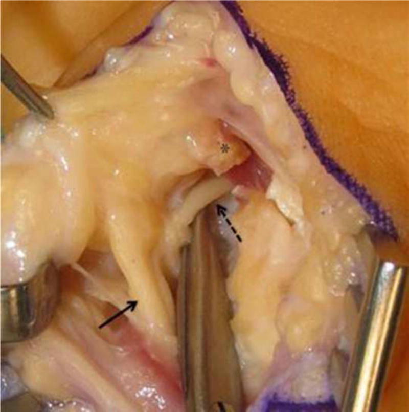
Magnified intraoperative photograph after pisohamate arch released. Shown are the ulnar nerve (solid arrow), the deep motor branch of the ulnar nerve (dotted arrow), and the stump of cut pisohamate arch (*).

Magnified intraoperative photograph after pisohamate arch released. Shown are the ulnar nerve (solid arrow), the deep motor branch of the ulnar nerve (dotted arrow), and the stump of cut pisohamate arch (*).